FMF Certification
Cervical assessment
Sonographic measurement of cervical length is useful in firstly, the prediction of pregnancies at high-risk of early preterm delivery, secondly, distinction between true and false labor in women presenting in threatened preterm labor, and thirdly, in the prediction of likelihood of cesarean section after induction of labor.
Requirements for certification
Please attend all online courses through our new FMF website.
From March 2026, registration will be required to qualify for access to your license extension for the risk calculation software. License files can be downloaded without audit from your own FMF page.
Further updates and requirements will be posted online soon.
Protocol for measurement
- The woman should have an empty bladder. She should be placed with her legs abducted to allow a full range of movements whilst scanning.
- Ultrasound transducer: 5MHz transvaginal probe. A disposable sheath should be used to cover the probe and the lubricating gel should be sterile.
- Gently place the probe in the anterior vaginal fornix to ensure a sagittal view of the cervix is obtained
- Identify the internal os, external os, cervical canal and endocervical mucosa. The endocervical mucosa should be used to define the level of the internal os. Care should be taken to distinguish between cervical canal and a thickened lower uterine segment coming together in the midline, which can give the false impression of a longer canal.
- Do not exert undue pressure on the cervix with the probe because this will falsely elongate the cervix.
- Magnify the picture so that the cervix occupies at least 75% of the image
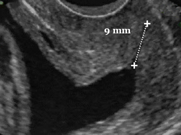
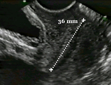
- Measure the distance between the internal and external os. Take 3 measurements (and pictures) over a period of about 3 minutes and record the best shortest measurement of the cervical length
- Note the possible presence of funnelling at the internal os. The endocervical mucosa will give an accurate definition of the amount of funnelling . Occasionally a thickened lower uterine segment can mimic a funnel and this can be identified by the absence of mucosa extending along the walls of the funnel
- Note the possible presence of dynamic changes in the cervix, defined by the appearance and disappearance of funnelling during the scan.
Vaginal scan: normal cervix
Vaginal scan: short cervix